In order to interpret your findings after collecting and preparing a urine specimen for microscopy you must obtain a high quality image. If you are not able to clearly see what you’re examining, it may be quite difficult to differentiate cell types and discern what inclusions are present within a cast.
Let’s take a look at two sets of images taken with the exact same microscope….
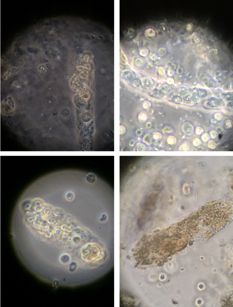
How can we take a better look at these urine sediments?
The key to improving image quality is attention to several important points:
- Ensure that the microscope objective, eyepiece, and condenser lenses are clean
- Ensure that the camera lens and adapter are clean
- Verify proper illumination of the specimen (Köhler Illumination)
- Choose the appropriate illumination modality
- Use glass slides and cover slips, not plastic multiple chambers slides
Here are a few tips:
- To ensure clear focus of all elements in the viewing field, the objects must all be in the same thin physical plane. This is best achieved by using a small drop of sediment and a large cover slip. Apply the cover slip carefully to avoid any air bubbles.
- Experiment with different illumination modalities (brightfield, darkfield, phase contrast, polarized) to find the one which best displays your findings.
- If you have enough urine, spin two tubes and prepare one for viewing unstained and the other using a Sternheimer-Malbin stain. Phase contrast is much better with unstained specimens whereas brightfield offers better resolution with stained specimens.
- Remember to use only high quality glass slides and cover slips and avoid using plastic multi chamber slides as the optical clarity is inferior and the thickness of the plastic prohibits proper use of oil immersion lenses.
- Use of the oil immersion lens provides a higher numerical aperture and higher resolution than can be achieved otherwise. I believe the highest resolution images are obtained with brightfield illumination of a stained specimen using an oil immersion objective lens.
Below are a few informative optical microscopy references:
Optical Microscopy
Immersion Oil & Refractive Index
6 Tips to Properly Clean Immersion Oil Off Your Objectives
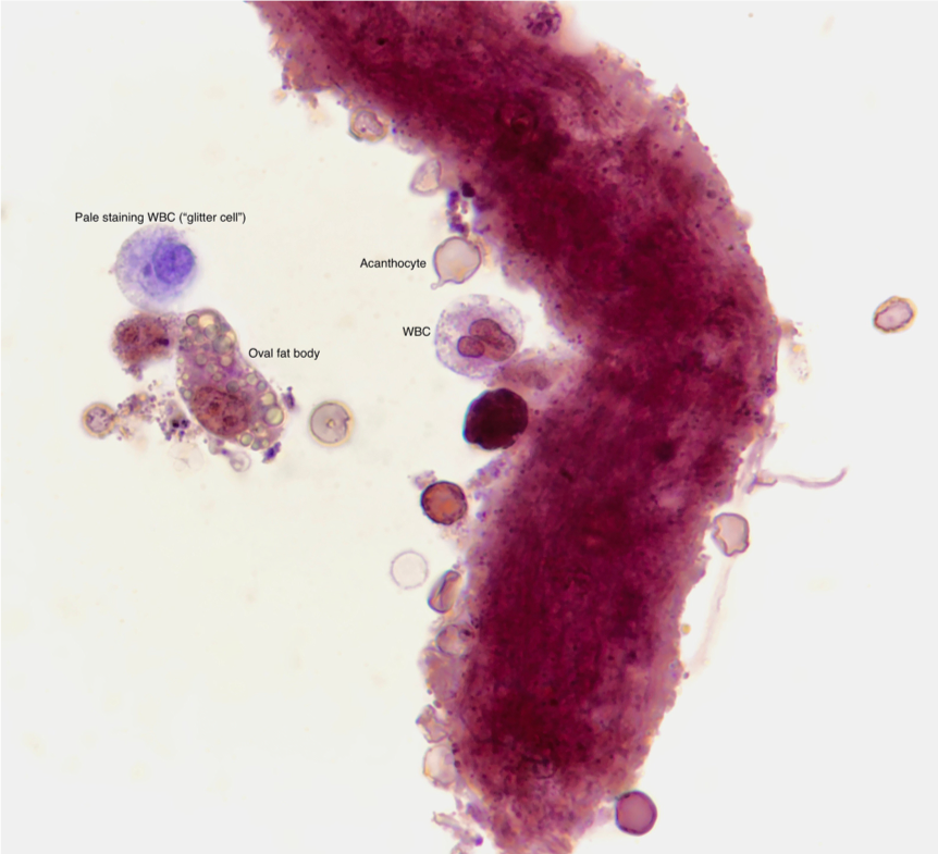
Below is a higher quality image, taken using the tips above. See the difference?
Post by: Jay Seltzer, MD